Skip to content

Retinal Detachment Surgery

There are several different surgery options for retinal detachment, depending on the severity, cause, and location.

What Is Retinal Detachment?

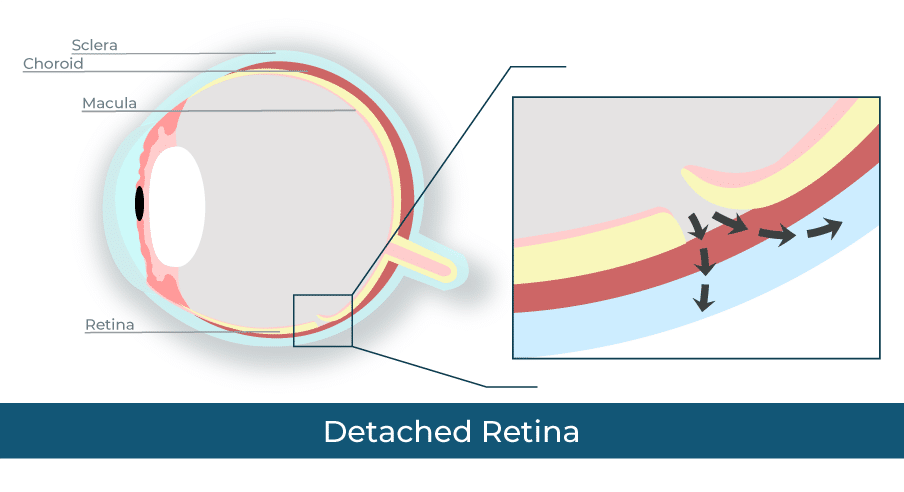
Retinal detachment is when the retina is pulled or torn out of position. A detached retina, if not treated promptly, can cause vision loss.

Your retina is the layer of cells at the back of your eye that detects light and turns that light into signals for your brain. These cells include photoreceptors, which give you color vision and night vision.

If the retina detaches, it doesn’t work properly.

Symptoms of retinal detachment include:

  • A gray curtain covering some of your field of vision
  • A shadow in your side vision
  • Seeing flashing lights
  • Seeing many new floaters

What are the Treatment Options for a Detached Retina?

There are five different surgical options for a detached retina. The type of treatment your doctor suggests will depend on how severe the damage is and what type of retinal detachment you have.

Freeze Treatment (Cryopexy)

Retinal cryopexy is a less invasive treatment option that a doctor may suggest if the tear in your retina is very small. It can often be done in the doctor’s office.

Your doctor will first put numbing medicine in your eye. Then they will take a small freezing probe and touch the white outside part of your eye, called the sclera, in the area right over the tear. The cold from the probe causes scar tissue to form around the tear, which holds the retina in place.

Laser Surgery (Photocoagulation)

Photocoagulation is a type of retina surgery that uses a medical laser. Like freeze treatment, your doctor may suggest it for small tears, and it can usually be done in a doctor’s office.

In laser surgery, your doctor will numb your eye, then shine a medical laser inside your eye. The laser makes small burns in your retina that create scar tissue around the hole. This seals the hole and holds the retina in place.

Pneumatic Retinopexy

In a pneumatic retinopexy, your doctor will use an air bubble to push your retina back into place. They then use freeze treatment or laser surgery to repair any tears in the retina.

To perform a pneumatic retinopexy, your doctor will first numb your eye. They’ll then insert a very small needle into your eye and remove a bit of fluid. Next, they insert a bit of air to replace that fluid. The air bubble will push your retina into place, and then your doctors will use freeze treatment or laser surgery as needed. The air bubble will disappear after a few days.

After surgery, you’ll need to keep your head in a certain position for several days to keep the bubble in place.

Scleral Buckle Surgery

In a scleral buckle surgery, your doctor will sew a small, flexible band to your sclera. This band presses on the sides of the eye, moving them inward. This helps your retina reattach.

This type of surgery is usually done under anesthesia. The band stays on your eye, but you won’t be able to see it.

Vitrectomy

A vitrectomy uses an air bubble to push the retina in place, like a pneumatic retinopexy, but the method is slightly different.

Your doctor will make small incisions in your eye. They’ll then use suction to remove the vitreous fluid. They replace the fluid with a bubble made of air, gas, or oil. This bubble pushes the retina back into place. Your eye will eventually replace the vitreous fluid you lost during surgery.

For several days or weeks after surgery, you’ll need to hold your head in a certain position to keep the bubble in place. You will also need to avoid flying in an airplane or climbing to high altitudes, as altitude changes will increase eye pressure.

Retina

Book Your Next Appointment Entirely Online.
Find An Appointment That Works For You!

What to Expect After Retinal Detachment Surgery

The recovery time for your retina surgery will depend on the specific surgery you had. Your doctor should give you an estimated recovery time, as well as what you may experience after. General things to expect after a retina surgery include:

  • Discomfort and pain: Your doctor will likely give you medication to manage this.
  • Eye patch: You’ll need to wear an eye patch to protect your eye. Your doctor will let you know how long you’ll need to wear it.
  • Floaters and flashing lights: It’s common to see these for a few weeks after surgery.
  • Rest: You’ll need to reduce your activity for a few weeks after surgery. Your doctor should tell you which activities are safe and which are not.

Risks of Retina Surgery

Retina surgery, like any type of surgery, has risks. These include:

  • Bleeding in the eye
  • Cataracts
  • The chance that the retina doesn’t properly attach or detaches again
  • Increased pressure in the eye, which can lead to glaucoma

While these risks can be scary, not treating a detached retina can cause you to lose your eyesight permanently.

Types of Retinal Detachment

Retinal detachment is categorized by cause. There are three types: Exudative, rhegmatogenous, and tractional.

Exudative Retinal Detachment

Exudative retinal detachment is caused by a build-up of fluid behind your retina that pushes the retina away from the back of your eye. This causes the retina to detach but doesn’t cause tears or breaks in the retina.
This fluid build-up is usually the result of leaking blood vessels or swelling. This can be caused by:

  • Age-related macular degeneration
  • Coats disease
  • Diseases that cause eye inflammation
  • Eye injury or trauma
  • Tumors in the eye

Rhegmatogenous Retinal Detachment

Rhegmatogenous retinal detachment is the most common kind of retinal detachment. It’s often the result of aging. As you age, the vitreous fluid, which is the jelly-like fluid inside your eye, begins to shrink. This shrinking can cause it to tug on your retina, tearing it. When the retina tears, vitreous fluid can get behind your retina and push on it, which detaches the retina.
Other causes of rhegmatogenous retinal detachment include eye injuries, eye surgery, and nearsightedness.

Tractional Retinal Detachment

Tractional retinal detachment is the result of scar tissue on the retina pulling the retina out of position. The most common cause of this is diabetic retinopathy, which can cause scarring on the retina. Other causes may include eye diseases or infections or swelling inside the eye.

Retina Surgery Centers in Arizona

We offer retina examinations and consultations across Metro Phoenix, Northern Arizona, and Southern Arizona. We want to make sure your experience is convenient and easy. Our eye surgeons also perform retina procedures at our own surgery centers. Learn more about our Ophthalmology locations.

Schedule an appointment online

Book Your Next Appointment Entirely Online.
Find An Appointment That Works For You!